COMPARATIVE NEUROANATOMY of SOCIAL SYSTEMS:
OXYTOCIN and VASOPRESSIN RECEPTORS
Oxytocin and vasopressin are two evolutionary-ancient hormones that are synthesized by the brain, and decades of research have now demonstrated that these hormones are potent modulators of social behavior in animals and humans. The overarching goal of my research program is to understand the neural mechanisms by which oxytocin and vasopressin affect mammalian social behavior, including humans and nonhuman animals. To tackle this comparative and translational work, it is important to first identify the target locations of oxytocin and vasopressin in the brain: namely, where in the brain are the receptors for these hormones? We use a variety of histological methods, such as receptor autoradiography and in situ hybridization, to identify the target locations of oxytocin and vasopressin in the brain.
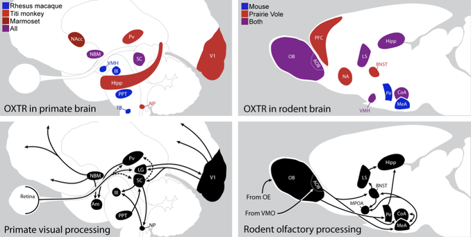
In the Freeman Lab, we perform this rigorous fundamental neuroanatomical work to characterize the underlying neural circuits of oxytocin and vasopressin's action, which can help identify neural targets for future, site-specific studies. I developed the first reliable method for the visualization of oxytocin receptors (OXTR) and vasopressin 1a receptors (AVPR1a) in primate brain tissue, which yielded the first map of the sites of action of these social hormones in non-rodent brains. By establishing the neural substrates that are responsible for the action of oxytocin in the brains of several primate species, my work guided future research into how oxytocin functions to modulate primate social behavior. I used this method to map oxytocin receptors across three primate species and found that they co-localize with brain areas that mediate visual attention and gaze direction. This pattern is distinct from that of rodent brains, where oxytocin receptors overlap with the circuits for olfactory processing and social memory. Thus, oxytocin appears to act in areas that process information from the sensory modality most heavily relied upon for navigating social environments: olfaction for rodents and vision for primates. By extending this work to the brains of other mammalian taxa, ongoing efforts in our lab will inform our understanding of the shared versus unique aspects of the neurobiology of sociality across species.
Freeman SM. Using Receptor Autoradiography to Visualize and Quantify Oxytocin and Vasopressin 1a Receptors in the Human and Nonhuman Primate Brain. Methods Mol Biol. 2022;2384:105-125. PMID: 34550571.
available here
Freeman SM, Young LJ. Comparative Perspectives on Oxytocin and Vasopressin Receptor Research in Rodents and Primates: Translational Implications. J Neuroendocrinol. 2016 Apr;28(4):10.1111/jne.12382. PMID: 26940141.
available here
Freeman SM, Inoue K, Smith AL, Goodman MM, Young LJ. The neuroanatomical distribution of oxytocin receptor binding and mRNA in the male rhesus macaque (Macaca mulatta). Psychoneuroendocrinology. 2014 Jul;45:128-41. PMID: 24845184.
available here
Freeman SM, Walum H, Inoue K, Smith AL, Goodman MM, Bales KL, Young LJ. Neuroanatomical distribution of oxytocin and vasopressin 1a receptors in the socially monogamous coppery titi monkey (Callicebus cupreus). Neuroscience. 2014 Jul 25;273:12-23. PMID: 24814726.
available here
THE OXYTOCIN SYSTEM of THE HUMAN BRAIN:
INVOLVEMENT in NEUROPSYCHIATRIC CONDITIONS
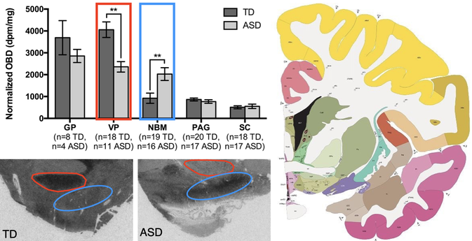
Due to the ability of oxytocin to enhance social behavior, it is now a promising therapeutic for the treatment of human psychiatric conditions that characterized by deficits in social function, such as autism spectrum disorder (ASD), schizophrenia (SZ), and social anxiety disorder. Indeed, oxytocin treatment can ameliorate some of the social symptoms of ASD and SZ, and genetic studies have implicated the oxytocin system in the etiology of both conditions. However, whether there are differences in oxytocin receptor expression in the brains of individuals with ASD compared to unaffected individuals is only now being investigated. The Freeman lab is working to determine whether there are underlying differences in the oxytocin receptor densities or locations in postmortem human brain tissue from donors with psychiatric diagnoses like ASD and SZ, compared to matched, unaffected specimens. After validating our method for the selective localization of oxytocin receptors in human brain tissue, we were the first to determine that individuals with ASD have dysregulated oxytocin receptor levels in two behaviorally relevant regions of the forebrain that are involved in visual attention (cholinergic basal forebrain) and reward/motivation (ventral pallidum). We have also recently shown that the dopaminergic pars compacta of the substantia nigra contains fewer cell-surface, mature oxytocin receptors in females with ASD compared to their unaffected female counterparts, as well as to males with ASD. Furthermore, a full characterization of the distribution of oxytocin and vasopressin 1a receptor binding and gene expression throughout the neurotypical human brain has not been performed, due to the mixed affinity issues described above. To this end, we have recently finished mapping oxytocin and vasopressin 1a receptors throughout the human brain. Once complete, this map can inform results from previous and future neuroimaging studies of the effects of intranasal oxytocin and vasopressin on human brain activity, and it can also enhance our understanding of the mechanisms by which oxytocin and vasopressin 1a receptors modulate human behavior.
Frehner SS, Dooley KT, Palumbo MC, Smith AL, Goodman MM, Bales KL, Freeman SM. Effect of sex and autism spectrum disorder on oxytocin receptor binding and mRNA expression in the dopaminergic pars compacta of the human substantia nigra. Philos Trans R Soc Lond B Biol Sci. 2022 Aug 29; 377 (1858): 20210118. PMID: 35858098.
available open access here
Freeman SM, Palumbo MC, Lawrence RH, Smith AL, Goodman MM, Bales KL. Effect of age and autism spectrum disorder on oxytocin receptor density in the human basal forebrain and midbrain. Transl Psychiatry. 2018 Dec 4;8(1):257. PMID: 30514927.
available open access here
Freeman SM, Smith AL, Goodman MM, Bales KL. Selective localization of oxytocin receptors and vasopressin 1a receptors in the human brainstem. Soc Neurosci. 2017 Apr;12(2):113-123. PMID: 26911439.
available here
Freeman SM, Frehner SS, Palumbo MC, Smith AL, Goodman MM, Bales KL (in prep). Characterization of the distribution of oxytocin receptor and vasopressin 1a receptor binding and mRNA throughout the neurotypical human brain.
BEHAVIORAL NEUROENDOCRINOLOGY of COYOTE SOCIAL BEHAVIOR
By studying the hormones, brains, and behavior of monogamous animals, we can glean unique insights into the biological mechanisms driving the formation and maintenance of long-lasting, selective social bonds. In the past several decades, this type of research has been conducted primarily in pair-bonding rodents, such as prairie voles. In our lab, we primarily focus on the coyote (Canis latrans) as a representative of the mammalian family Canidae, in which all wild species of canid have been shown to exhibit a monogamous mating system, at least in some environments. We have ongoing projects to study coyote partner preference behavior, proximity maintenance between pairs (including co-sleeping), genetics and pharmacology of the oxytocin & vasopressin systems int the coyote, and olfactory communication and social recognition. This work takes place at the Utah Field Station of the USDA National Wildlife Research Center, in collaboration with Dr. Dustin Ranglack and other local coyote experts, Drs. Julie Young and Eric Gese.
Turano A, Brummer SP, Young JK, Freeman SM. Can a traditional partner preference test quantify monogamous behavior in captive coyotes? Behav Processes. 2023 Jan 21;206:104832. PMID: 36693577.
available open access here
Freeman SM, Adamis S, Anderson T, Ihrig H, Johnson N, Measom M, Nielson B, Rich M, Staheli A, Webb M (in prep). Oxytocin receptor and vasopressin 1a receptor distribution in the brain of the coyote (Canis latrans).
SOCIAL VISUAL ATTENTION in MONOGAMOUS MONKEYS
Following our discovery that oxytocin receptors in the primate brain tend to overlap with circuits governing visual attention, I was eager to develop methods to study social looking behavior in monogamous primates. To this end, I have continued collaborating with my postdoc mentor Dr. Karen Bales at the California National Primate Research Center (Davis, CA), where we use non-invasive eye-tracking technology to study the visual social behavior of the monogamous South American primate: the coppery titi monkey (Plecturocebus cupreus). We hope to expand this work to include eye-tracking research in the closely-related, but nocturnal, owl monkey (Aotus sp.), through collaborations with the Keeling Center for Comparative Medicine and Research (Bastrop, TX).
Ryan AM, Freeman SM, Murai T, Lau AR, Palumbo MC, Hogrefe CE, Bales KL, Bauman MD. Non-invasive Eye Tracking Methods for New World and Old World Monkeys. Front Behav Neurosci. 2019 Mar 5;13:39. PMID: 30890923.
available open access here
Lau AR, He L, Loyant L, Baxter A, Bauman MD, Bales KL, Freeman SM (in prep). Non-invasive eye tracking reveals partner-directed looking behavior in monogamous titi monkeys (Plecturocebus cupreus).
